Effects of <33™ technology on wound healing as shown in two clinical wound healing trials performed under the guidance of Dr. Dirk Lange, Director of Basic Science Research at the Stone Centre at Vancouver General Hospital and Associate Professor in the Department of Urologic Sciences at the University of British Columbia, Faculty of Medicine.
Wound Healing
The results from these wound healing studies demonstrate that exposure to <33™ Wound Healing Formula Frequency Plates (“<33”) accelerates the third phase of wound healing and helps speed up the growth of new tissue.
The phase involving the formation of functional networks of small blood vessels with perfusion of red blood cells (neovascularization) required for healing was shorter in wounded skin exposed to <33™ devices indicating that healing of these wounds was accelerated resulting in the more rapid growth of fresh tissue and formation of a new layer of outer protective tissue.
Wounds that were not exposed to <33™ devices showed a higher degree of neovascularization and fibrosis indicating that the tissue damage is still greater in these wounds after 14 days.
Study 2 – Results & Discussion.
Our results indicate that <33™ accelerates Stage 3 of wound healing.
The results from this wound healing study demonstrate that exposure to <33™ accelerates the third phase of wound healing and helps speed up the growth of new tissue. We observed that the levels of neovascularization (the regeneration of blood vessels across the damaged tissue) and fibrosis (the growth of fresh connective tissue triggered by the fibroblasts) were higher for longer periods of time in wounds that were not exposed to natural frequencies compared to those that were exposed to the natural frequencies. While one may think that these processes should be enhanced in wounds that are healing more rapidly, it is in fact the opposite. The prolonged period of higher degrees of neovascularization and fibrosis in the wounds that were not exposed to the <33 indicates that the tissue damage is still greater in these wounds compared to those that were exposed to <33 and that it took longer activity for these wounds to heal. In other words, a longer degree of healing activity is required to reach the same healing as indicated by the similar degree of healing observed in Day 14. This is also evident by the existence of more organized tissue in the wounds exposed to <33, which is another indication of advance healing. Visual examination and images taken show that the incisions exposed to the <33™ devices are healing faster than those that are not exposed to the technology as was observed in the prior Study 1. The microscopy analysis of cross-sections of the wounded areas show that Phase 3 of the four- phase healing process is the one most effected by the presence of <33™ devices. The microscopy images of the cross- sections of the biopsies are shown in Figures 1 and 2.
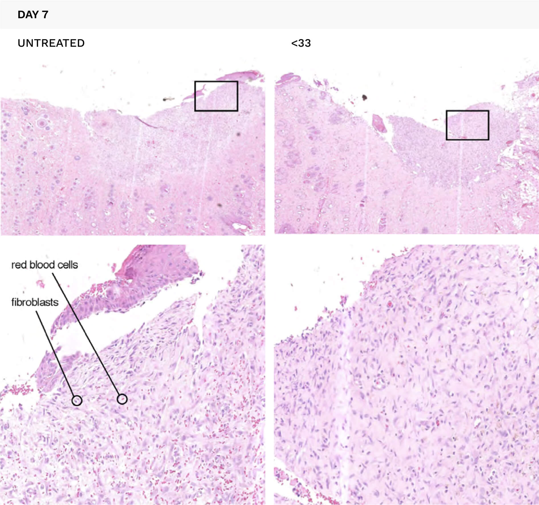
Figure 1. Microscopy images of a representative cross-section of the wounded area on Day 7. The bottom images are enlargements of the inserted areas highlighted by the black rectangles. Red blood cells are shown in red and fibroblast cells are stained purple. Lower level of neovascularization in the exposed wounds is indicated by fewer red blood cells while less neovascularization is indicated by less purple stained cells.
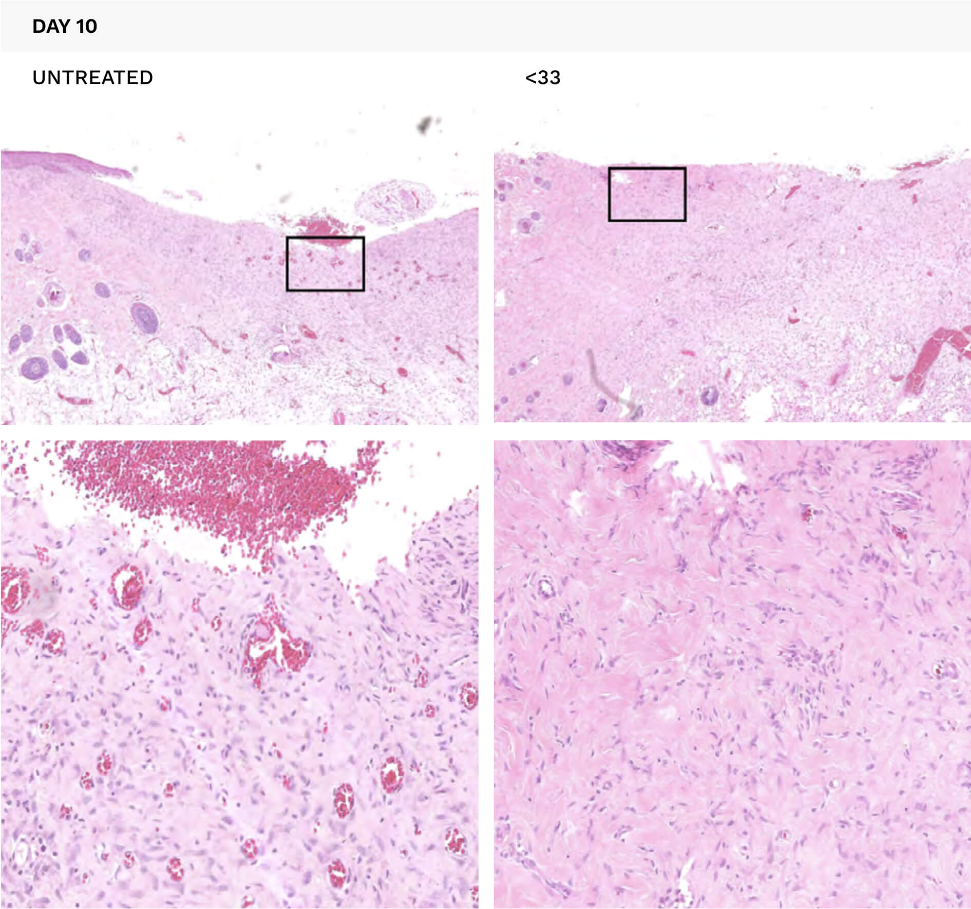
Figure 2. Microscopy images of a representative cross-section of the wounded area on Day 10. The bottom images are enlargements of the inserted areas highlighted by the black rectangles. Red blood cells are shown in red and fibroblast cells are stained purple. Lower level of neovascularization in the exposed wounds is indicated by fewer red blood cells while less neovascularization is indicated by less purple stained cells.
The loss of neovascularization is indicated by significantly less of the red blood cells in Figures 1 and 2, while the maturation of the fibrotic component is indicated by the decrease in purple cells. These analyses indicate that the levels of neovascularization and fibrosis in the later stage of the wound healing process were higher in the untreated compared to the treated wounds. The significance of these findings to wound healing is that both neovascularization and fibrosis suggest the presence of active ulcers, which can slow the healing process.
The levels of neovascularization and fibrosis can be assessed and compared by giving each a numerical score based on the observations from the microscopy studies. When these values are compared (by subtracting the values of the untreated group from those from the <33 group), a direct comparison can be made. Both neovascularization and fibrosis are higher in the untreated group compared to the <33 treated group.
The fact that we observed lower levels in the <33™ treated wounds exposed to <33™ Wound Healing Formula Frequency Plates demonstrates an effect on wound healing.
The studies were conducted under the guidance of Dr. Dirk Lange, Director of Basic Science Research at the Stone Centre at Vancouver General Hospital and Assistant Professor in the Department of Urologic Sciences at the University of British Columbia, Faculty of Medicine.
Clinical trials and studies shown have all been carried out with the highest level of scientific rigour at some of the best equipped, independent facilities, universities, and laboratories available.
If you’re interested in further details about the studies, please contact us. The signing of a non-disclosure and confidentially agreement may be required.